flow cytometry results for lymphoma
In most cases the lineage can be identified as T-cell B-cell or myeloid and a. A locked padlock or https means youve safely connected to the gov website.

Use Of Flow Cytometry In The Phenotypic Diagnosis Of Hodgkin S Lymphoma Grewal 2019 Cytometry Part B Clinical Cytometry Wiley Online Library
Immunophenotyping by flow cytometry is a sensitive method for detecting residual or recurrent disease in the peripheral blood of patients with an established diagnosis.

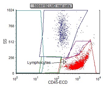
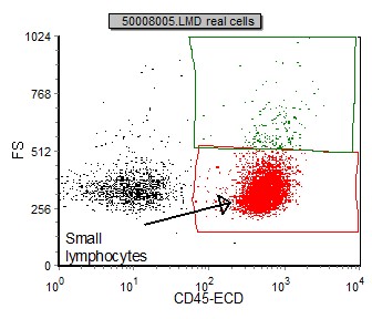
. Nonhematologic malignancy can be suspected if less than 75 percent of the cells show CD45 common leukocyte antigen. Clinical Diagnosis of Classical Hodgkin Lymphoma by Flow Cytometry. After review of the clinical history and morphology a panel of markers is selected for each case by a board-certified hematopathologist.
Immunophenotyping Flow Cytometry for Hematolymphoid Neoplasia. It is used to detect abnormal hematolymphoid populations determine what cell surface markers they express and integrate immunophenotypic findings with morphologic and. Correlation of grade of lymphoma with flow cytometric CD19 forward scatter.
Grade 1 follicular lymphomas had a percentage of cells at or beyond the 500-channel mark ranging from 012 to 66 median 46 whereas grade 2 follicular lymphomas had a percentage ranging from 412 to 1255 median 7. These results will explain if any abnormal cells are present and what types of cells they are as a part of your diagnosis. 88185 - Each Remaining Marker.
FL cells are surface Ig heavy and light chain - predominantly IgM. Results are reported by Aspirus Reference Laboratory in 2-4 working days. Click here for instructions on how to download the free FCS Express Reader to view and manipulate the sample cases.
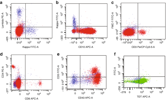
Flow results cannot be used alone to diagnose malignancy. Flow cytometry is rapid and appears to be virtually diagnostic of non-Hodgkins lymphoma when a majority of cells are B cells with an abnormal kappalambda ratio 40 or 025. It is part of Flow Cytometry in Clinical Diagnosis by John.
Flow cytometry is an important test that confirms the diagnosis of CLL by checking a persons blood cells or bone marrow for signs of. Lineage identification can provide a confirmatory diagnosis or differential diagnosis prognosis and treatment options. Therefore flow cytometry is an important integral part of lymphoma diagnosis even in.
The advantages of flow cytometry are based largely on its ability to analyse rapidly and simultaneously multiple cell properties in a quantitative manner. It is a highly aggressive lymphoma that is usually found in extranodal sites or presenting as an acute leukemia. Depending upon flow cytometry immunophenotyping results a healthcare practitioner may determine how likely your cancer will respond to treatment and how aggressive the treatment might be.
Activation of GSK-3β in CHL results in inhibition of the Wntβ-catenin signaling cascade and its abnormal accumulation in the nuclei of both Reed-Sternberg cells and Hodgkin cells 65. Cancer Answer Line 8662238100. The example report is intended to be a basis for further discussion within the flow cytometry cominunity on whether minimum reporting standards for leukemia andor lymphoma flow cytometry results can and should be developed.
The advantages of flow cytometry are based largely on its ability to analyse rapidly and simultaneously multiple cell properties in a quantitative manner. Flow cytometry has become an important tool in the diagnosis of mature lymphoid neoplasms and the determination of prognosis in selected cases. This test generates a hematopathology report with a diagnosis and interpretation of findings.
They express bcl-2 CD10 CD19. B-Lymphoblastic LeukemiaLymphoma BCR-ABL1-like 2016 WHO new provisional entity. Professor and Medical Director of Flow Cytometry Mayo Clinic Rochester MN 2019 MFMER slide-2 Flow Cytometry in Acute Leukemias.
Mayo Clinic Mayo Medical Laboratory On-line information. Flow cytometric immunophenotyping is. This translocation has been associated with the development of Follicular lymphoma.
The genetic hallmark of BL is a reciprocal translocation of the MYC gene on chromosome 8 most commonly. A translocation between chromosome 14 and 18 results in the overexpression of. Share sensitive information only on official secure websites.
Other results Differential count. Burkitt lymphoma BL a highly aggressive B-cell lymphoma represents approximately 25 of all non-Hodgkin lymphomas NHLs. Flow cytometry is a laser-based technique used to detect and analyze the chemical and physical characteristics of cells or particles.
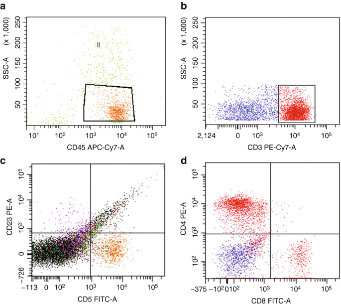
Flow cytometric leukemia and lymphoma analysis may aid in identifying the tumor lineage for diagnostic and prognostic purposes. Ad Be Seen by Mayo Clinics World-Class Lymphoma Experts. Discover the Latest Transplant Treatment Options at Mayo Clinic.
Results showed that in 916 56 percent the diagnosis of lymphoma or cancer could be suspected by flow cytometry alone while 416 were consistent with the final tissue diagnosis of normal or reactive hyperplasia. Flow cytometry also helps to define lymphoma subgroups for example the distinction between CLL hairy cell leukemia or other subgroups. Lymphoma Immunophenotyping by Flow Cytometry.
The white blood cell differential takes a closer look at your white blood cells and the percentage they make up in your blood. FLOW CYTOMETRY FOR HODGKINS LYMPHOMA DIAGNOSIS. Download a Treatment Guide.
Available online at. LeukemiaLymphoma Immunophenotyping by Flow Cytometry. The interval of time receipt of sample at Mayo Clinic Laboratories to results available taking into account standard setup days and weekends.
This test is usually done after atypical results are seen on a complete blood count or white blood cell WBC differential. The first day is the time that it. Leukemia and lymphoma analysis by flow cytometry aids in identifying the tumor lineage which in most cases is identified as T cell B cell or myeloid.
5 segs 52 lymphocytes 32 monocytes 9 eosinophils. Typical ranges are. Flow cytometry has become an important tool in the diagnosis of mature lymphoid neoplasms and the determination of prognosis in selected cases.
88184 - 1st Marker. Results may be delayed or the sample rejected if pertinent andor required information is conflicting or missing. Burkitts lymphoma BL is a cancer of the lymphatic system in particular B lymphocytes.
It is most commonly used to evaluate bone marrow peripheral blood and other fluids in your body. Flow cytometry laboratory medicine leukemia lymphoma phenotypes human. Hodgkins disease cannot be detected by flow.
However flow cytometry results usually make certain lymphoma entities extremely likely and others very unlikely. O 1995 Wiley-Liss Inc. BL preferentially involves extranodal sites such as the small intestine or jaw or may manifest as acute leukemia.
This flow cytometry test is used to diagnose leukemia or lymphoma. Flow Cytometry Lymphoma Immunophenotyping. Detection of a population of cells expressing CD38 and CD138 in the peripheral blood is useful in establishing a diagnosis of plasma cell leukemia when used in conjunction with.
With increasingly rare exceptions diagnosis of lymphoproliferative diseases and myeloid stem cell disorders relies on a multimodal analysis in which flow cytometry plays a significant role While a detailed technical overview of flow cytometry is beyond the scope of this paper and can be. See Flow Cytometry Report from Aspirus Reference Laboratory. 88187 88188 88189 - Professional.
Flow cytometry as a tool for the diagnosis of CHL has not been useful in the past due to the difficulty in.
Mantle Cell Lymphoma Mcl Flow Cytometry
Flow Cytometry Of Mature And Immature T Cell Lymphoma Springerlink
International Clinical Cytometry Society
Clonality Assessment And Detection Of Clonal Diversity In Classic Hodgkin Lymphoma By Next Generation Sequencing Of Immunoglobulin Gene Rearrangements Modern Pathology

Selected Flow Cytometric Immunophenotyping Plots From Fine Needle Download Scientific Diagram

Examples Of Cd200 Expression In Mantle Cell Lymphoma By Flow Cytometry Download Scientific Diagram

Hodgkin S Disease Ask Hematologist Understand Hematology Non Hodgkins Lymphoma Hodgkins Lymphoma Non Hodgkin
Flow Cytometry Of Mature And Immature T Cell Lymphoma Springerlink
Follicular Lymphoma Fl Flow Cytometry

Use Of Flow Cytometry In The Phenotypic Diagnosis Of Hodgkin S Lymphoma Grewal 2019 Cytometry Part B Clinical Cytometry Wiley Online Library
Diffuse Large B Cell Lymphoma Dlbcl Flow Cytometry

Summary Of Flow Cytometry Immunophenotypic Results For Anaplastic Large Download Table

Flow Cytometric Immunophenotyping Performed On The Same Plasmablastic Download Scientific Diagram

Flow Cytometric Presentation Of A Large B Cell Lymphoma A Forward Download Scientific Diagram

Anti Brdu Antibody Clone Bu20a Bio Rad Analysis Expressions Map Screenshot

Demystifying The Diagnosis And Classification Of Lymphoma A Guide To The Hematopathologist S Galaxy Mdedge Hematology And Oncolo Diagnosis Lymphoma Oncology

Forward Scatter Fsc Vs Side Scatter Ssc Plots After Doublet Download Scientific Diagram
Flow Cytometry Of Mature And Immature T Cell Lymphoma Springerlink

Flow Cytometry Results Of Suspected Central Nervous System Download Scientific Diagram